 |
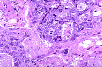
44.A mitotic figure is seen in the
center, surrounded by a poorly differentiated squamous cell carcinoma with pleomorphic
cells and minimal pink keratinization. In general, mitoses are more likely to be seen in
malignant neoplasms. Remember, though, that normally cells are actively dividing in bone
marrow, gonads, and gastrointestinal tract.
44.图示中央有一核分裂相,周围见形态不一的低分化鳞癌细胞和很少的角化。一般来说,恶性肿瘤的核分裂相多见,但记住,骨髓,生殖腺和胃肠道的细胞分裂活跃。 |